Prendre
rendez-vous

Sesamoid injuries

Métatarsalgie

What is it?

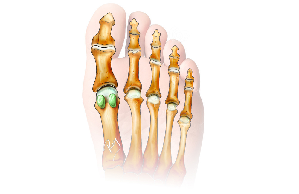
Anatomically, a sesamoid bone is a bone located in a muscle or tendon. Its etymology comes from the Latin “sesamum”, which means sesame, given their resemblance to a sesame seed.

They are located in several areas of the human body such as the patella (kneecap) or the thumb in the hand.

In the foot, this type of injury is encountered in many forms, but generally speaking it refers to the two small bones beneath the first metatarsal head, which are defined as “sesamoid”.

Lésions des sésamoïdes
  • Fig. 1

    Sesamoid bone located under the head of the first metatarsal.

They have several functions in the foot:

  • – Shock absorption
  • – Role in supporting the body’s weight
  • – Act as a pulley

Shock absorption and their role in bearing the body’s weight largely explain the susceptibility of these structures to direct or chronic trauma. However, their pulley function increases the effect of the various flexor muscles on which they depend and for which they are used as insertions.

There are several types of sesamoid injuries, which generally differ by their occurrence or radiological images. The main injuries are:

Sesamoiditis (30%)

Inflammatory damage generally caused by chronic overload.


Stress fracture/fatigue (40%)

It typically appears following prolonged activity (running, marathon, etc.) or with the development of chronic sesamoiditis.


Acute fracture (10%)

As a result of axial trauma, for example (a fall, etc.).


Arthritis (<10%)

Often associated with hallux valgus or diffuse arthritis of the big toe.

Clinical presentation

Whether chronic or acute, sesamoid injuries always present as a pain under the big toe next to the affected sesamoid. Mobilising the toe can increase the pain. Finally, the presence of thickened skin at the lesion (= hyperkeratosis) is usually an indication of a chronic process.

Sport is often a source of significant pain, especially when running or doing endurance sports. It is worth highlighting the situations that trigger pain and the aggravating factors.

The clinical assessment is always accompanied by radiological images in order to highlight, for example, a fracture. The tests are often accompanied by a scan or an MRI to clarify any uncertainties. They can also be used to diagnose the viability of a sesamoid bone after a fracture.

When should you consult a specialist?

When chronic pain or traumatic pain presents under the big toe, it is advisable to consult a doctor to avoid the development of a chronic condition or a worsening of the injury.

Do you have any questions or would you like to see a specialist?

MAKE AN APPOINTMENT
Click here